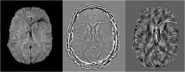
FORT DETRICK, Md. (Aug. 6, 2012) -- A new dimension in imaging technology detects minute levels of vascular damage in the form of bleeding, clots and reduced levels of oxygenation that may better illuminate our understanding of brain injury, particularly related to trauma.
Currently, the U.S. Army Medical Research and Materiel Command's Telemedicine and Advanced Technology Research Center is managing a related project that is being led by Dr. E. Mark Haacke of Wayne State University.
Haacke recently presented his work in susceptibility weighted imaging and mapping, or SWIM, to a national panel of military and civilian medical experts. In this current project, he is exploring advanced magnetic resonance imaging methods and SWIM to improve diagnosis and outcome prediction of mild traumatic brain injury.
"This study is just one example of the promising research that TATRC supports. Collaborations among the investigators we bring together may lead to creative solutions we hadn't imagined," said Col. Karl Friedl, TATRC director.
In 1997, Haacke's team developed susceptibility weighted imaging, a highly sensitive technique to detect the presence of blood products. According to Haacke, it has been proved to be the most sensitive approach to visualizing cerebral microbleeds and shearing of vessels in traumatic brain injury, or TBI.
"These conditions do seem to be reliable indicators of injury because we have imaged hundreds of adults over the years, of all ages, and rarely find them in the normal control population," said Haacke.
In recent years, Haacke's team and other neuroimaging researchers have applied concepts similar to SWIM to provide a new measure of iron content through quantitative susceptibility mapping. Haacke's approach, SWIM, is a rapid method that not only provides a quantitative map of iron but at the same time reveals the presence of cerebral microbleeds and abnormal veins.
Iron in the form of deoxyhemoglobin can also be used to measure changes in local oxygen saturation, important for evaluating potential changes in local blood flow or tissue function (similar to what is seen in stroke using SWI). SWIM can also be used to monitor changes in iron content over time to see if previous iron deposition is being resorbed or if bleeding continues, both important diagnostic pieces of information for the clinician.
"SWIM is among the highest quality and fastest types of quantitative susceptibility mapping," said Haacke. "We believe it could be in much wider use in about a year."
Haacke has been working with researchers throughout the world for more than five years applying his techniques specifically to traumatic brain injury, stroke, Parkinson's disease and multiple sclerosis.
In this current project, he has demonstrated that there is a lower impact load, either inertia or direct impact forces, which may damage only veins, and he has shown medullary vein damage that has not been visualized with other techniques. The medullary veins drain the frontal white matter of the brain, so reduced blood flow here could possibly impair the higher level frontal neurocognitive functions. In light of this, treatments that improve blood flow to the brain might be a promising direction to pursue.
While many investigators have focused on arterial changes related to brain injuries, Haacke has remained focused on the veins.
"Veins have relatively more fragile vessel walls than arteries and are more susceptible to damage during head injury," said Haacke. "This important component of the vascular system is often overlooked but may help us better diagnose what is wrong."
"Doctor Haacke's team has a different slant for studying these injury regions that may lead to a new avenue in diagnosis and treatment for traumatic or other types of brain injury," explained Dr. Anthony Pacifico, who manages TATRC's Medical Imaging Technologies Portfolio.
"For instance, the study of dementia could well benefit from SWI and SWIM," said Haacke. "Perhaps as much as one-third of all dementia is vascular dementia."
Haacke and Dr. Zhifeng Kou are working to complete a larger database of normal and mildly brain-injured imaging scans and define the appropriate parameters so that SWIM can be run at most clinical sites.
TATRC manages leading-edge research in various areas of medicine and technology, seeking synergies among projects that may quickly advance new products into the field to improve the care of the nation's warfighters.
Related Links:
VIDEO: Traumatic Brain Injury roundtable raises awareness
PEO Soldier: New Gen II Helmet Sensor will Provide Data to Help Detect Traumatic Brain Injury
Army, NFL collaborate on traumatic brain injury helmet sensors
'Pockets of excellence' across Army, but work still needs to be done on health of force
STAND-TO! Traumatic Brain Injury
Defense Centers of Excellence for Psychological Health & Traumatic Brain Injury
Telemedicine & Advanced Technology Research Center
U.S. Army Medical Research and Materiel Command
Defense Centers of Excellence for Psychological Health & Traumatic Brain Injury on Facebook

Social Sharing